The University Cancer Center's Breast Care Program has been on the cutting edge of advances in both the screening and treating of breast cancer, earning accreditation by the American College of Radiology as a breast imaging center of excellence.
In 2014, 239,109 people in the United States were diagnosed, according to the Centers for Disease Control and Prevention’s webpage on breast cancer. In the same year, 41,676 people died from breast cancer.
Breast cancer is typically detected by mammograms, which are X-ray images of the breast. Mammography reduces the risk of dying of breast cancer by around 40 percent. As technology improves, those numbers will continue to grow, said Jennifer Harvey, division director of breast imaging and co-director of the University Cancer Center’s Breast Care Program.
The recommended start age for women to get mammograms varies, but Harvey said that women should begin getting annual testing at the age of 40.
“Although [breast cancer is] less common for women in their 40s, it tends to get aggressive,” Harvey said. “Because of that, we really do need to find it early. Only about 25 percent [of breast cancers] are undiagnosed under the age of 50, but they account for about a third of breast cancer deaths. It certainly is less common but certainly not rare.”
Harvey said that as long as women are in good health, they should continue to receive annual mammograms.
At the University Medical Center, women have two options for their mammograms — 2D or 3D.
Traditionally, mammograms are 2D, like standard X-rays, and are available widely throughout the country. The Cancer Center implemented 3D mammograms over five years ago, and now, over half of examinations use this technology.
“Probably about 60 percent of the mammograms we do now are 3D, also called tomosynthesis,” Harvey said. “Instead of a single image of the breast, a machine takes 11 to 15 low dose X-rays at different angles over the breast and those are reformatted to 1 millimeter [image] slices.”
These slices allow radiologists to look at the various levels of the breast to spot any abnormalities, rather than just a single, comprehensive image.
By providing a more in-depth look into breasts, 3D mammograms finds around 30 percent more instances of breast cancer. Often, those cancers identified tend to be characterized as invasive types that are in danger of spreading to lymph nodes under the arms, Harvey said.
In addition, the typical 2D mammography results in 10 to 12 percent of women being asked to return because of potential abnormality on the image, according to Harvey. Extra pictures and an ultrasound can determine whether an abnormality actually exists, but 3D mammography limits the need for this step, meaning that almost a third less women get called for extra pictures.
Because most women over the age of 40 receive annual mammograms, the Cancer Center does 60 to 100 screening examinations daily.
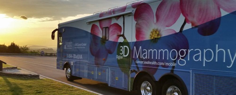
One way the Cancer Center reaches more women is through a mobile mammography bus which is capable of performing 25 mammograms daily, according to the Cancer Center’s webpage.
Another improvement to breast cancer screenings has recently been implemented at the University Health System’s Mammography Center Northridge, which has begun to use screening ultrasounds.
“To my knowledge no one else in this area has the technology,” Harvey said.
The new machine, installed in the last month, uses sound waves to look for breast cancers rather than X-rays. This allows women who have dense breast tissue to get more accurate results.
“When we add ultrasound to a mammogram, we can find about 30 percent more cancers for women with dense breast tissue,” Harvey said. “Ultrasound cancers are dark on white tissue so we can see cancers on ultrasound that we can't see on mammography [where they show up as white on white tissue].”
Though the screening ultrasound technology was implemented in the last few weeks, other testing measures have been used by the University Health System for much longer.
The Cancer Center’s High-Risk Breast and Ovarian Cancer Clinic has been working with patients for the past 15 years. The clinic determines risk based on family history and genetic testing, which further sets the University apart from other medical centers.
“Most hospitals don't have a high risk clinic,” Harvey said. “[Our practitioners] are great at figuring out if somebody is at risk, and if so what kind of imaging and other tests they may need.”
If screening detects that a person has breast cancer, there are two options — attempting to save the breast through breast conserving therapy or removing the entire breast in a mastectomy.
Standard breast conserving therapy has three components.
“The first component is an operation where we remove the tumor from the breast ... typically called a lumpectomy,” David Brenin, chief of breast surgery and co-director of the University Breast Care Program, said.
The second component consists most commonly of a sentinel lymph node biopsy, which checks to see if the cancer has spread under the arm to the lymph nodes.
Once the tissues heal from surgery, the patient undergoes the third component — radiation therapy on the breast.
Whole breast radiation is the standard method of radiation therapy and requires patients to receive treatment for a few minutes a day at a radiation facility. The process takes between three-and-a-half weeks to six-and-a-half weeks.
A separate option for radiation therapy is intraoperative radiation therapy, and is fairly unique to the University.
“We can actually give all the radiation during surgery,” Brenin said. “With two brief operations the patient's treatment is completed with a total time of two hours.”
Intraoperative radiation therapy is available at a few locations throughout the state, though the exact treatment varies among centers.
“[Other centers are] using a technique that we believe is inferior to what we're doing now,” Brenin said. “We have a special way of doing it that we believe is going to be shown to be better.”
One of the University’s newest studies into treatment options starts Friday and looks into ultrasound ablation combined with immunotherapy.
“[The treatment is] using ultrasound waves to ablate, or heat up, the breast cancer in the breast or lymph nodes underneath the arms and cause a local immune response,” Brenin said. “We’re going to ramp up that immune response with a drug [that] ... tells white blood cells to attack tumor cells.”
Breast cancer typically does not elicit a significant immune response from the body on its own, so the treatment attempts to help increase the body’s response through focused ultrasound and medication.
The University is working to better treat breast cancer, especially for more advanced stages. This study hopes to help accomplish that.
“For patients with stage four breast cancer unfortunately the prognosis is not great,” Brenin said. “We’re starting to investigate at UVa and elsewhere new treatments ... to improve our ability to treat patients with advanced stage breast cancer.”
Patients diagnosed with stages one or two have a better outlook, according to Brenin.
“[For] patients with stage one breast cancer, more than 95 percent of them will be alive in five years,” Brenin said. “With stage two, more than 85 percent will be alive in five years. The prognosis for breast cancer has improved greatly over the past 10 years.”
Improvements in detection and treatment of breast cancer have led to these results, Brenin said, and the University looks to further improve on them for the future.


