“A snail mail, regular mail letter was sent addressed to Mr. James Ryan … President of U.Va., and in this three page letter was a paper check for $1 million dollars,” said Michael Wiener, a researcher in the department of Molecular Physiology and Biological Physics at the School of Medicine.
Early January, Wiener won a $1 million grant from the William M. Keck Foundation to use distances within a protein to map its structure. This new method promises to be cheaper, faster and easier than traditional techniques. Wiener was the University’s nominee for the Keck Foundation Grant in the Medical Science category. The press release announcing this award was published Feb. 15.
If the new method proves effective, it could be utilized by the greater scientific community to study biological structures and aid in the development of pharmaceuticals. This grant spans over three years and is almost completely matched by the University.
The Keck Foundation is an independent philanthropic organization based in Southern California and is famous to researchers for funding “high-risk, high-reward” scientific projects. According to Joel Baumgart — the senior research program officer in the Office of the Vice President for Research — most research grants in the University are funded by federal agencies such as the National Institutes of Health. However, these agencies tend to give money to research that already has a lot of preliminary data and is the next logical step in that researcher’s work.
“[Wiener’s idea] is a little bit too risky for the canonical, federal funding agencies,” Baumgart said. “That's where the foundations like Keck come in. They look for the space between what the agencies will fund.”
Although Wiener is the first researcher from the University’s School of Medicine to receive a Keck Foundation grant, in 2016 University biologist Eyleen O’Rourke was awarded $1.2 million from the same foundation. Baumgart credits some of these recent funding acquisitions to the expansion of the University’s corporate and foundations relations office.
“What we learned is there's an ecosystem to research,” Baumgart said. “The idea alone, the idea in a box or in a drawer, is not enough itself to carry the day. What you need is ... the foundations office.”
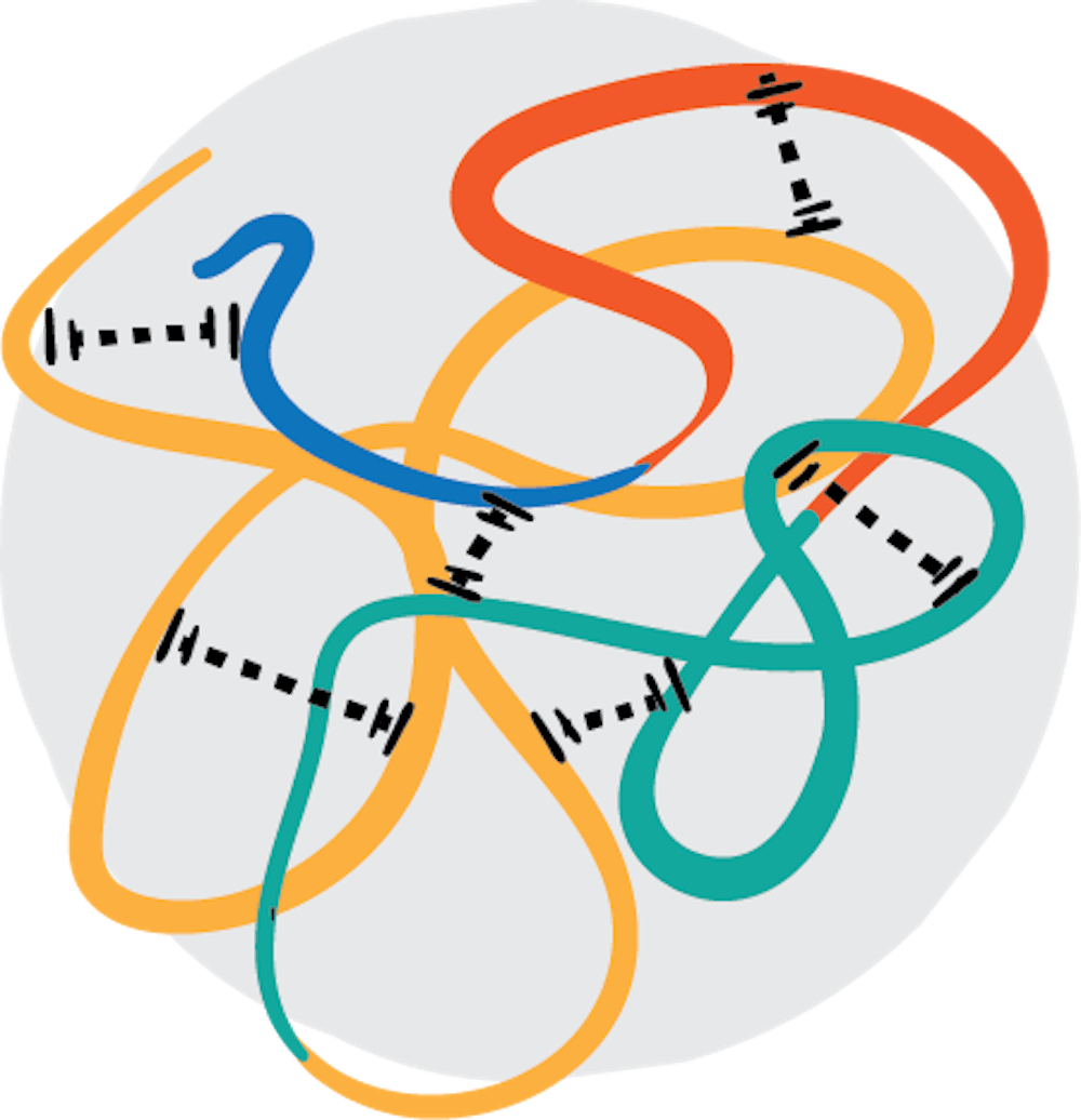
A focus of Wiener’s research is preparing numerous, lower-resolution samples of proteins — rather than risky and time-consuming samples — to solve the shape of a molecule. Wiener explained that determining the shape of a molecule is like creating an image. One method is taking a picture of the the whole — which in the submicroscopic realm is expensive and often does not work — and another method is finding distances between the parts of the whole and using those distances to create an image.
“Instead of one sample that has all the goodies, you make a large number of samples that will be much easier to make — and fast and cheap,” Wiener said. “Each one of them gives you a small bit of information which you can then assemble together to get the entire structure.”
Other methods used to solve protein structures are x-ray crystallography, in which a crystallized protein sample is exposed to x-ray beams, as well as cryo-electron microscopy, which cools samples to extremely low temperatures and studies them with an electron microscope. Wiener’s proposed method — called serial solution scattering structure determination, or S4D — uses a large number of samples with chemical labels that are analyzed with x-rays and computational methods to determine the structure of a protein.
This technique could also save time and resources needed to prepare samples for analysis. According to Brandon Goblirsch, a research associate in Wiener’s lab, when studying the structure of molecules, the majority of time spent in the lab is preparing samples.
“S4D is really … targeting something that is a real bottleneck to structural biology and biological sciences,” Goblirsch said. “Just preparing samples, and doing it in a cost-effective and efficient way, frankly, [is] costly in terms of reagents but also people’s time and career time.”
Another aspect of S4D is the possibility of determining structures in a cell-like environment. Lukas Tamm, chairman of the Molecular Physiology and Biological Physics department in the University’s Medical School, explained that proteins, especially ones embedded in the membrane of a cell, are notoriously difficult to map because other components are attached to them. Once isolated for study, the protein is no longer in its natural system.
“In the living cells, they’re in more of a natural environment,” Tamm said. “And so they have all kinds of other components bound to them that you would see in a living cell that you would perhaps purify away when you isolate them and put them in a crystal.”
The next immediate steps for Wiener’s research are preparing the samples using small proteins called peptides and adding labeling agents to them. Sample preparation will occur in Wiener’s lab — in the University’s Fontaine Research Park — and in the next few months the researchers will take the labelled proteins to the Department of Energy’s National Synchrotron Light Source II, a facility in the Brookhaven National Laboratory in New York that houses beamlines that will analyze the sample and provide necessary data to evaluate Wiener’s method.
Once Wiener’s research team obtains data points for their samples from NSLS-II, that data will be analyzed by Wiener’s collaborators — Ken Dill, a computational scientist from Stony Brook University, and Lei Wang, a professor at the University of California, San Francisco, who will enable S4D to be used in living cells.
“I will be doing the research for all three years of this grant,” Wiener said. “My collaborators … will start in year two. So essentially we're driving it forward to get results and certain core experiments done, which will then set the stage to dial in these other technological developments.”
According to Goblirsch, Dill will take the experimental data obtained from NSLS-II and use it to reconstruct three-dimensional models of proteins.
“Molecules are three-dimensional — they move [and] breathe,” Goblirsch said. “Ultimately we want to know the three-dimensional architecture so one way is to … get lots of these distances, but then somebody has to develop software and the algorithms and the know-how to feed that into a three-dimensional model that takes advantage of all those distances.”
According to Wiener, the chemical and technological techniques necessary for labeling these proteins in live cells will be a challenge to this project.
“I would say that they are technological challenges, rather than conceptual challenges,” he said.
The money from the grant will be used to buy scientific instrumentation and support scientists and technicians in his lab and in the labs of his collaborators. He plans to stay in contact with his collaborators through Skype and Collab.
Baumgart explained that federal grants for research provide an additional 61 percent of the amount of each grant to the University to pay for general facilities, such as electricity and WiFi. Grants from foundations such as the Keck do not pay the extra 61 percent, and because of this, the University in the past tended not to encourage applications for foundation grants. However, Baumgart suggests that after obtaining a large grant from a foundation, more federal grants tend to follow.
“Somebody who wins a Keck [is going to] get federal grants,” said Baumgart.
Wiener has had this idea for solving protein structures for at least a decade. Long-term, he plans for this project to be a large part of his research focus for the rest of his career.
“The driver for my going into research science has been the notion that science at its best is a creative endeavor,” Wiener said. “It's a craft. It's an art. It's my art.”


