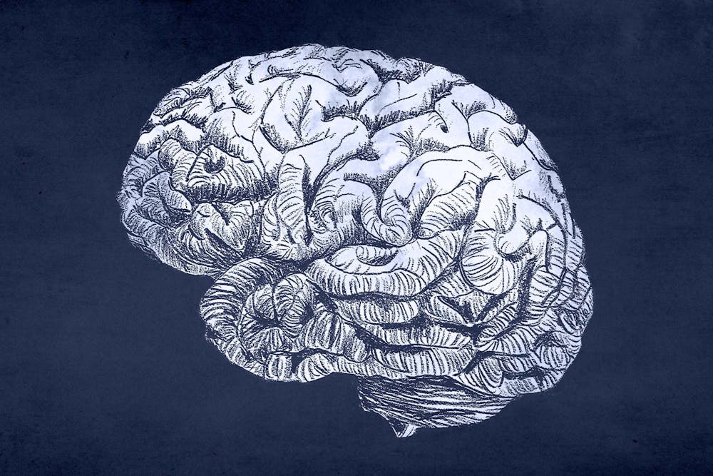
The University recently launched a study regarding the effects of long-term blast exposures on the brain health of military service members.
The research team includes principal investigator James Stone, vice chairman of research and associate professor of radiology and imaging, as well as a team of researchers at the National Institutes of Health, Walter Reed Army Institute of Research and the Naval Medical Research Center. They aim to apply their research to blast exposure mitigation, prevention and therapy rehabilitation.
Participants of the study include service members who experienced repetitive low-level exposure — meaning who were repeatedly in close proximity to an explosive during detonation — that may lead to manifestation of issues within the brain over time, Stone said.
Examples of people who fit these criteria include service members who utilize explosives to break through hard structures, explosive ordnance disposal personnel that clear roadside bombs and explosives and individuals who work with heavy weapons such as mortars.
“They may be exposed to many, many of these exposures over a career as a function [of] what they do day to day,” Stone said.
To better understand the effects that service members face, Stone researches questions regarding the magnitude, frequency, overall length of time and sometimes cumulative exposure that feeds into the adverse impact that blast encounters have on the brains of service members.
According to Stone, this study is part of an overall broader phased process of understanding the cause and effect relationship between blast exposures and an individual’s physical response.
“[Phase one includes] defining the phenomena … or what may be altered by the blast,” Stone said.
Phase two, often occurring in conjunction with phase one, describes the relationship between the phenomena and the magnitude, frequency and overall cumulative exposure to blasts. The study will apply conclusions drawn from the first two phases to inform how military training and operations should be conducted to mitigate and prevent brain injuries precipitated by blast exposures and therefore protect service members.
To collect the data needed for each of the different phases, the researchers utilize the resources of the University’s medical imaging research facilities, located within the department of radiology at the School of Medicine to conduct their research, said Dr. Kiel Neumann, assistant professor of radiology and medical imaging. They use research dedicated to magnetic resonance imaging, positron emission tomography and computed tomography equipment to complete their work.
The MRI machines demonstrate how different areas of the brain are connected and allow researchers to study the overall activity of the service members’ brains.
“When it comes to magnetic resonance imaging, we're able to use certain elements of magnetic resonance imaging to give us a very high resolution look at what the brain cortex looks like versus what the brain white matter pattern looks like,” Stone said.
This high-resolution cortical imaging helps determine whether the service members’ total amount of brain cortex is changing over time, which is an indication of brain injury. Additionally, the MRI allows researchers to study the movement of water within the brain to identify white matter injury, or injury to the inner tissue of the brain, that may result from repetitive low level blast exposure.
The study’s methodology also includes a technique that combines the manufacturing of molecular imaging agents with an imaging modality known as Positron Emission Tomography.
“It’s been hypothesized that blasts may engender some forms of lasting or not lasting neural inflammation within the brain,” Neumann said.
The molecular imaging agent manufactured in the study, specifically DPA 714, binds to cells that present the inflammation. According to Neumann, there are two comparisons being made with the molecular imaging agents — an intraindividual comparison and an interindividual comparison.
“The intraindividual comparison … would be comparing areas that don't show any signal to areas that do show signal [within one person],” Neumann said. “We attribute the areas that show signal to be wholly derived from the presence of the imaging agent being in that part of the brain.”
In contrast, interindividual comparisons consider the same areas of the brain of subjects who have been exposed to blasts versus control subjects who haven't been exposed to blasts.
“In theory, [interindividual comparison subjects] should not show the same signals within their brain,” Neumann said.
The intraindividual comparison studies the areas in the brain of an individual that produce a signal versus areas of the brain that do not. Signaling is determined by whether or not the imaging agent is present in an area of the brain. Because the agent binds to cells experiencing inflammation, the presence of a signal indicates inflammation. In contrast, the interindividual comparison studies the presence, or lack thereof, of neuroinflammation in control subjects with subjects who have been exposed.
“[The study also correlates] between structure and function to be able to understand what the overall context of any of [the study’s] objective findings may be,” Stone said.
The study uses neurocognitive or neuropsychological measures such as specific questionnaires to understand the cognitive or psychological manifestations of brain imaging evaluations.
Stone also indicates that previous research serves as the foundations of this study. One 2014 article, “The effects of low level blast exposure on the nervous system: is there really a controversy?” — cowritten by Dr. Gregory A Elder, research professor at Icahn School of Medicine at Mount Sinai, and Stone — demonstrates the correlation between previous and current studies.
“I think that the basic questions we were asking back in 2014, we're still in large part trying to answer today even though we know more of the pieces of the bundle now,” Elder said.


