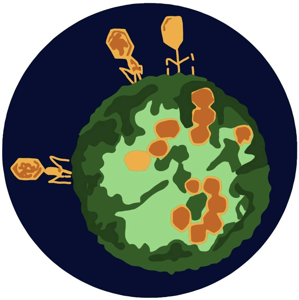
A team of researchers at the University School of Medicine recently published an article in the Journal of Biological Chemistry about their study of HIV fusion to cell membranes using cryo-electron microscopy. The study determined the initial steps of HIV infection in cells — information that can be applied to understanding the infection process of other viruses including SARS-CoV-2, the virus that leads to COVID-19 infection.
Previous studies revealed that the HIV infection process includes the fusion of viral membranes to cell membranes. This is achieved by binding viral surface proteins, or proteins located on the surface of a virus, to receptor proteins, located on cell membranes.
According to Amanda Ward, medical student in the Tamm Lab, the binding of the membranes of viruses to cells is a process that is largely the same even when considering a diverse group of viruses.
Specifically for HIV, spike proteins found on the surface of the virus for binding are similar to the spike proteins found on influenza, Ebola and SARS-CoV-2 viruses, said Lukas Tamm, director of the University's Center for Membrane and Cell Physiology.
According to Ward, while the exact proteins used in the fusion of HIV versus viruses such as influenza vary, the steps for fusion are similar. Therefore, having a technique that can observe the slight variations in the initial steps of viral infection can help attain an understanding of the overall initial infection process and enable further research.
To identify the specific intermediate steps of HIV fusion and understand how they are inhibited, researchers utilized techniques known as cryo-electron tomography and cryo-electron microscopy.
Cryo-electron tomography allows researchers to get “snapshots as an enveloped virus undergoes membrane fusion [which depict the] first steps of how [HIV] gets into a cell and can establish infection,” Ward said.
Cryo-electron microscopy is a specific form of electron microscopy, Ward said.
Electron microscopy works the same way as light microscopy but instead of shining a light at the sample and observing its interaction with the light, it introduces electrons to the sample and is performed in a vacuum so that as soon as an electron contacts anything solid it shatters, Ward said.
The cryo-electron microscopy technique won the 2017 Nobel Prize in Chemistry and is useful for sample preparation and data processing. In the University’s study, this technique was used to collect and freeze very thin samples of HIV and “blebs,” or mini cells so rapidly that the water within the sample does not form a crystalline structure as seen in ice, said Ganser-Pornillos, assistant professor of molecular physiology and biological physics at the School of Medicine.
The benefit behind using blebs is that “[whole] cells are too large to be frozen that quickly, [so] we pinched off small pieces of membranes from those cells [to form blebs] that are small enough ... to be subjected to rapid freezing,” Tamm said.
With Cryo-electron microscopy you can see exactly what is going on when the virus interacts with the cell without any stain or artifacts to disrupt the image, Ganser-Pornillos said. This contrasts traditional electron microscopy, which requires staining samples and therefore disrupts the structure of the samples themselves.
According to Ward, the study’s findings revealed the normal process of HIV fusion and how it is disrupted by the newly-discovered proteins Serinc3 and Serinc5. These proteins are able to block HIV infection and may provide a new avenue for designing therapeutics that could inhibit HIV fusion to cells.
Additionally, experimental results suggest that Serinc3 and Serinc5 inhibit the overall fusion of HIV particles without targeting a specific step in the fusion process.
Because previous research indicates that energy is required for each step of the fusion pathway, the study’s paper suggests it is plausible that Serinc3 and Serinc5 increase the energy needed to advance through different intermediate fusion steps. This additional energy requirement would inhibit HIV infection.
Ward said that in response to this study’s findings, she would like to conduct further research on how the viral membrane itself could be important to the fusion process.
“The end goal here is to have a detailed mechanism of how viruses infect cells, so that means understanding how every molecule rearranges at every step of this process ... This is one step on the way to that,” Ward said.


